Microscopy Images
Bile nephrosis occurs in patients with liver failure, and is associated with a clinical history of jaundice and elevated total bilirubin levels, usually greater than 20 mg/dL. Patients can present with acute kidney injury (AKI) or chronic kidney disease.
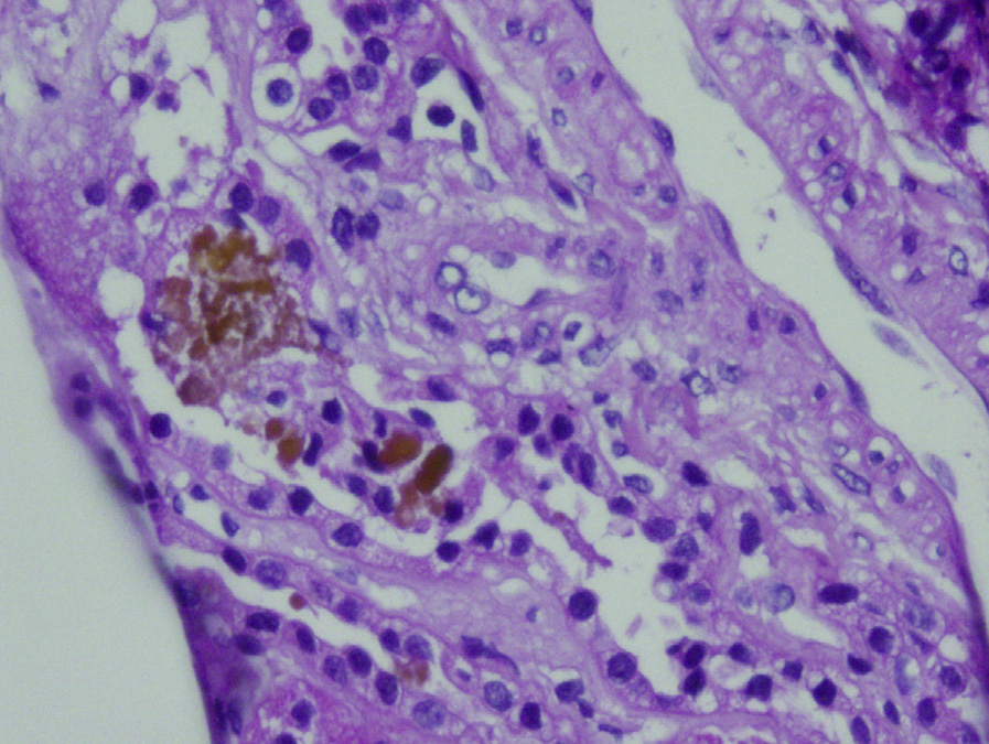
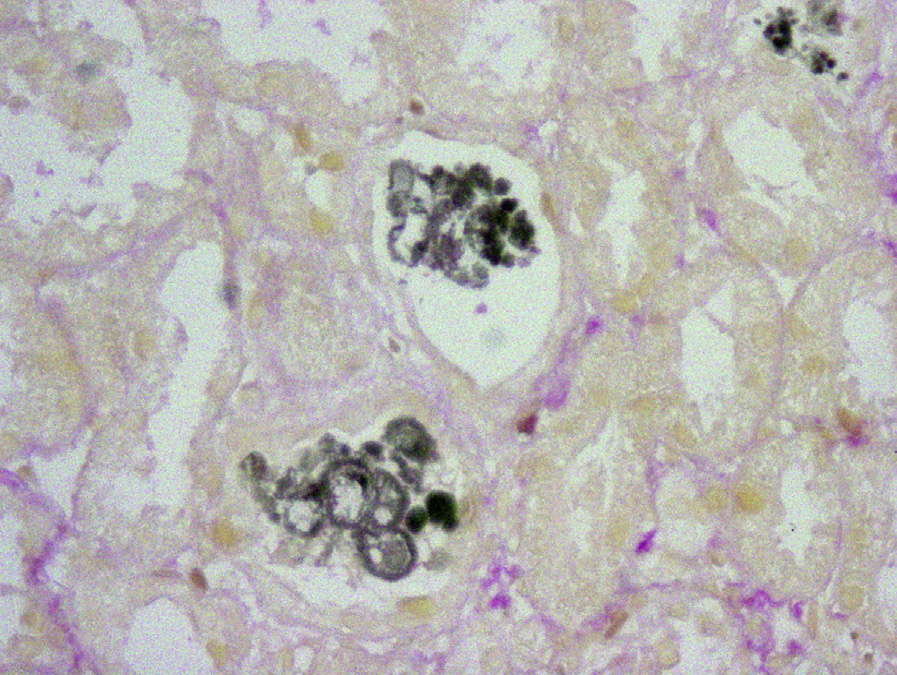
Light microscopy: Intratubular yellow-green granular casts are present predominantly in distal tubules and collecting ducts, with associated acute tubular injury with intratubular cellular debris. Casts are dark green by Hall stain.
Immunofluorescence microscopy: Noncontributory.
Electron microscopy: Noncontributory.
The etiology is multifactorial. Hepatorenal syndrome may be present, resulting in decreased kidney perfusion and ischemic tubular injury. This injury is reversed if the kidney is transplanted into a recipient without liver disease. Furthermore, the bilirubin and bile salts composing the casts are directly toxic to tubular epithelium. Distal tubule and collecting duct obstruction by casts composed of bile components and Tamm-Horsfall (uromodulin) proteins also contribute to AKI.
Acute tubular injury (eg, due to ischemia) can have granular casts related to cellular debris; however, these cases lack bile-stained casts, and Hall stain gives negative results. Myoglobin cast nephropathy has granular to globular reddish casts in distal tubules and collecting ducts that are positive by myoglobin immunostain. Hemolysis-associated hemoglobin casts are red, granular, and stain for iron (with Prussian blue). They are usually located in distal tubules and collecting ducts. Light chain cast nephropathy has intratubular casts that show metachromasia with fractured appearance and syncytial giant cell reaction, with monoclonal light chain staining by immunofluorescence microscopy.