Microscopy Images
Collapsing glomerulopathy is associated with marked proteinuria, rapid decrease in kidney function, and a poor prognosis. This lesion is unresponsive to corticosteroid therapy alone. There is a strong preponderance in African Americans versus whites with collapsing glomerulopathy.
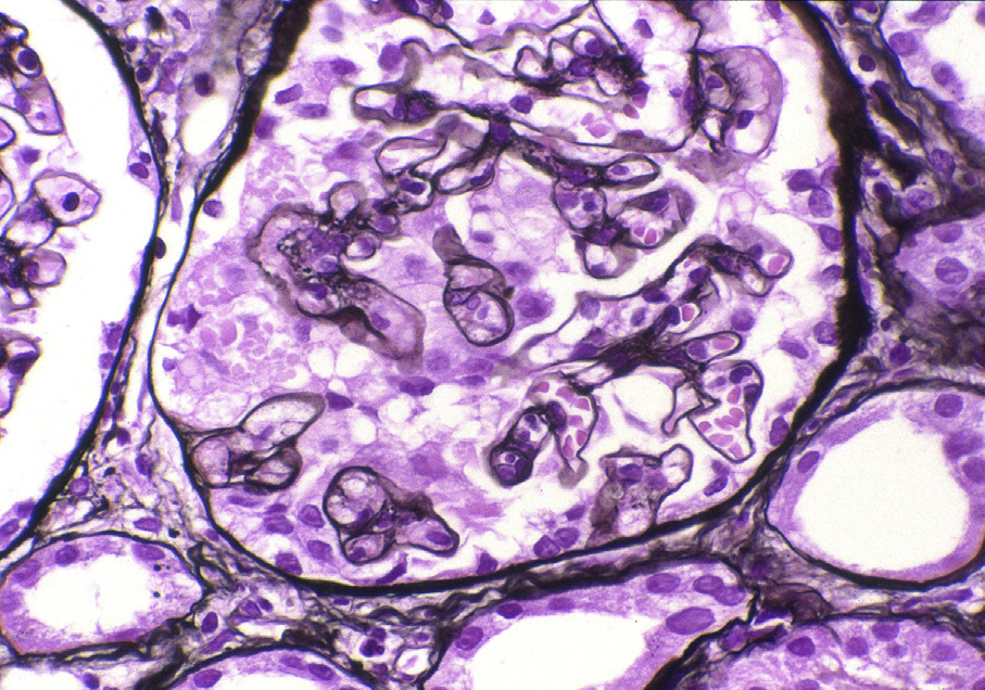
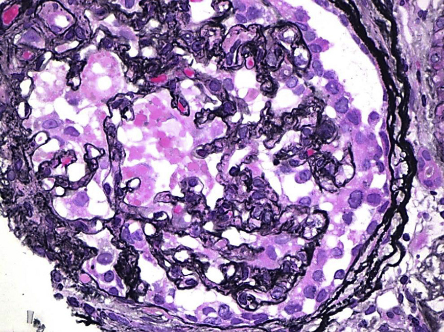
Light microscopy: Segmental or global glomerular tuft collapse with overlying visceral epithelial hyperplasia and hypertrophy. Hypertrophied visceral epithelial cells have frequent protein droplets.
Immunofluorescence microscopy: No or limited deposits (nonspecific immunoglobulin M and C3 staining in collapsed segments).
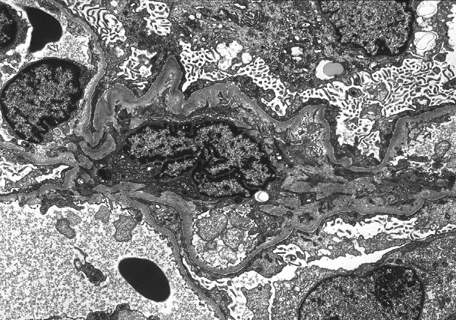
Electron microscopy: Glomerular basement membranes are wrinkled and collapsed. Overlying visceral epithelial cells show hypertrophy and hyperplasia, with frequent vacuoles and protein droplets. There is extensive foot process effacement and no or limited mesangial deposits. HIV (human immunodeficiency virus)–associated nephropathy or lupus nephritis– associated collapsing lesions show reticular aggregates, in contrast to idiopathic collapsing glomerulopathy, no reticular aggregates are present.
The etiology of collapsing glomerulopathy has not yet been discovered. There is proliferation and dedifferentiation of mature podocytes with loss of cyclindependent kinase inhibitor 1B (p27Kip1) expression in areas of collapse. Viral agents (HIV and parvovirus) have been proposed as a possible etiology. Treatment with pamidronate or calcineurin inhibitors has been linked to collapsing glomerulopathy. Patients with a risk allele variant of the apolipoprotein APOL1, common in African Americans, have an increased incidence of collapsing glomerulopathy.
Crescentic glomerulonephritis has parietal epithelial proliferation, but generally does not have reabsorption droplets. Crescents also often show fibrinoid necrosis and glomerular basement membrane breaks, features that distinguish them from collapsing lesions.
Secondary collapsing glomerulopathy can be related to infection (eg, HIV and parvovirus), drugs (eg, bisphosphonate and calcineurin inhibitors), severe vascular disease (eg, thrombotic microangiopathy and cocaine use), and autoimmune disease (eg, systemic lupus erythematosus).