Microscopy Images


Diffuse mesangial sclerosis presents with nephrotic syndrome at birth or within the first year of life. It may occur as part of Denys-Drash syndrome or as an isolated lesion. Patients with Denys-Drash syndrome typically are 46XY with ambiguous or female external genitalia, and streak (undeveloped) gonads or testes. Progressive kidney disease develops, usually by age 4 years. Patients have increased risk for Wilms tumor.
Light microscopy: Glomeruli are small and condensed in appearance, with early lesions showing increased loose mesangial collagen that progress to sclerosis with dense collagen without hypercellularity.
Podocytes do not show hyperplasia but may be immature and cobblestone-like.
Immunofluorescence microscopy: No specific staining.
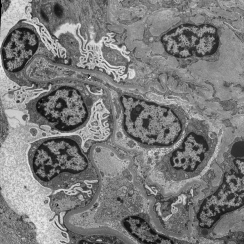
Electron microscopy: Extensive foot process effacement without deposits, but increased collagen within mesangial areas.
Patients with either isolated or syndromal forms of diffuse mesangial sclerosis have mutations in the WT1 gene, which encodes a transcription factor.
Tubules may occasionally be dilated when extensive sclerosis develops, but this is not an early or dominant feature, as seen in congenital nephrotic syndrome of Finnish type. Congenital nephrotic syndrome of Finnish type is also distinguished by lack of dense collagenous material within the mesangium.