Microscopy Images
Glomerulonephritis with dominant C3, a form of C3 glomerulopathy, typically presents with subnephrotic proteinuria and hematuria. Although a minority of patients may present with nephrotic syndrome, about half present with hypertension and impaired glomerular filtration rate. Up to 15% progress to end-stage renal disease. C3 glomerulopathy is rare with a typical age of onset of 30 years (range, 7-70 years).
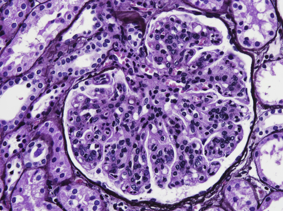
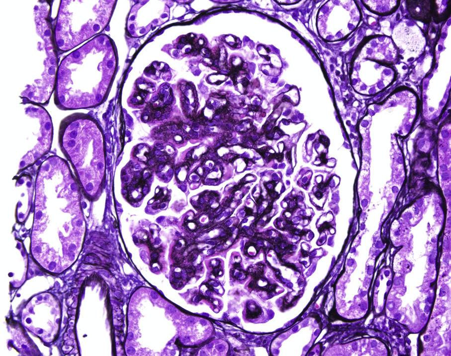
Light microscopy: Most biopsies show a membranoproliferative pattern. Other less common histologic patterns include mesangial proliferative, diffuse endocapillary hypercellularity, and crescentic.
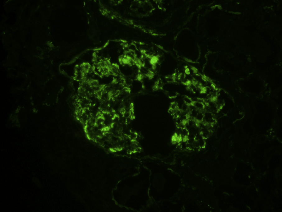
Immunofluorescence microscopy: Dominant or only C3 mesangial and capillary loop staining, at least 21 stronger than immunoglobulin and C1q staining.
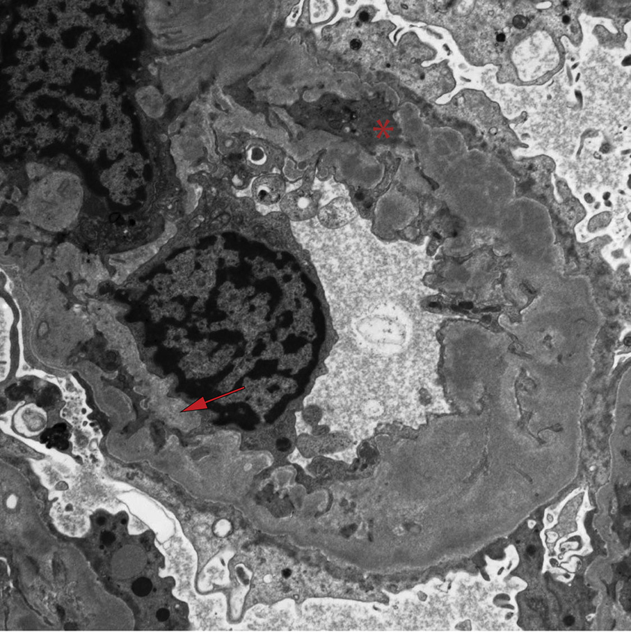
Electron microscopy: Mesangial and subendothelial deposits with occasional subepithelial deposits, possibly hump-like, without dense transformation of the deposits.
Abnormalities in alternative complement pathway regulation, genetic or acquired.
Dense deposit disease can present with similar light microscopy and immunofluorescence findings, but has diagnostic ribbon-like dense deposits within the glomerular basement membrane, not seen in this form of C3 glomerulopathy. Postinfectious glomerulonephritis can present with a similar pattern of C3 dominant staining by immunofluorescence; however, endocapillary hypercellularity with numerous neutrophils and subepithelial hump-like deposits revealed by electron microscopy are features that help distinguish it from glomerulonephritis with dominant C3.
Some patients with pathologic features indistinguishable from typical postinfectious glomerulonephritis have an atypical prolonged clinical course, and may have underlying complement dysregulation, indicative of a C3 glomerulopathy.