Microscopy Images
Source: AJKD Atlas of Renal Pathology II (atlas series 2015–2025); original article published 2025. original article record.
This page preserves the historical source-atlas wording. It is an educational reference, not a diagnostic or treatment recommendation, and has not been independently medically reviewed. Interpret morphology together with clinical, laboratory, serologic, genetic, and other relevant data.
Patients with lysozyme-associated nephropathy are usually aged 65-78 years, with male preponderance. Most patients present with acute kidney injury or acute kidney injury superimposed upon chronic kidney disease. A minority of the patients show partial Fanconi syndrome. Serum lysozyme levels are typically elevated in patients with lysozyme-associated nephropathy.
The most common etiologies for lysozyme-associated nephropathy are chronic myelomonocytic leukemia, acute myeloid leukemia, rare cases of metastatic neuroendocrine carcinoma, myelofibrosis, or sarcoidosis. In a minority of patients an underlying disease process may not be identified.
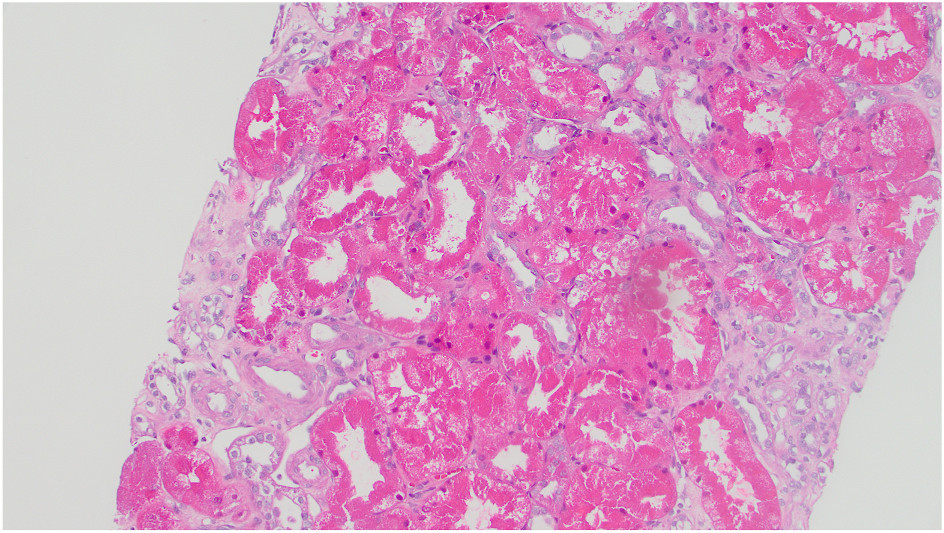
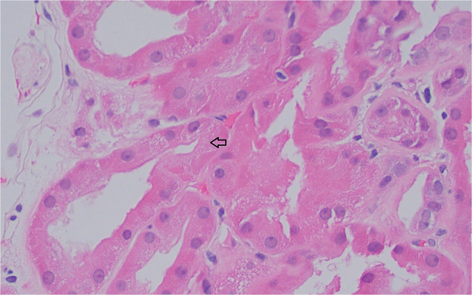
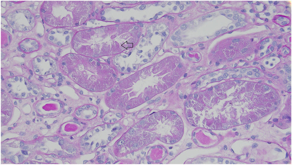
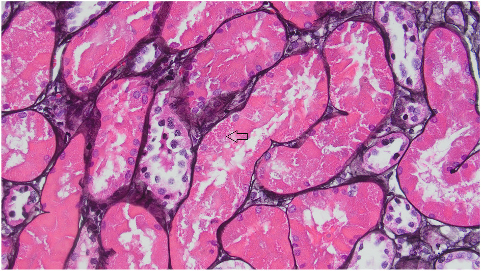
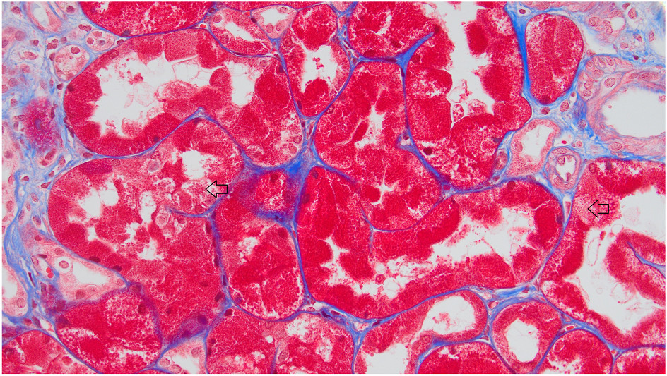
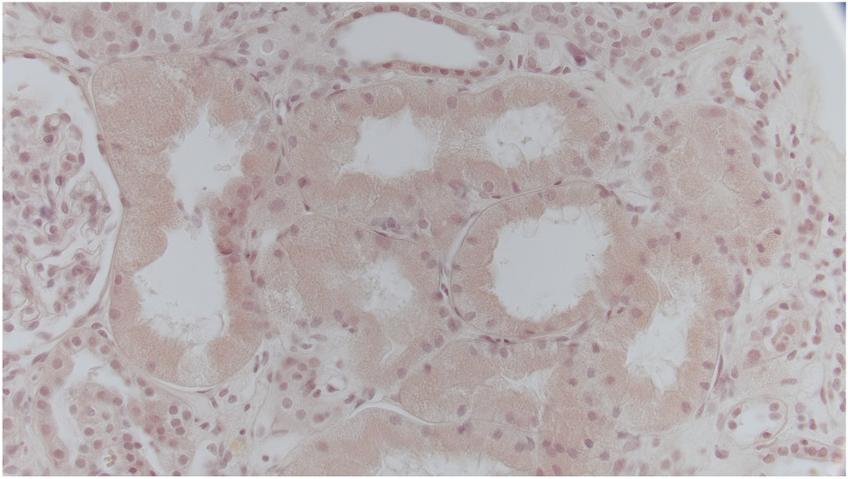
Light microscopy: Lysozyme-associated nephropathy is characterized by swelling of proximal tubular cells by cytoplasmic hypereosinophilic inclusions, which are usually pale on periodic acid–Schiff (PAS) stain and negative on Jones methenamine silver. They may demonstrate weak, pale, nonpolarizing staining with a Congo red stain and appear bright red (fuchsinophilic) on trichrome stain.
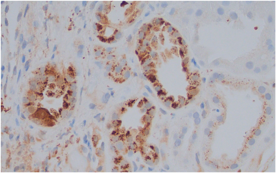
Immunohistochemistry for lysozyme shows characteristic diffuse intracytoplasmic inclusion staining, which is distinct from normal scattered background staining of proximal tubules. Tubules show acute injurious changes, including loss of brush border and cytoplasmic blebbing, and can show concurrent chronic injuries of tubular atrophy with associated interstitial fibrosis. Some case reports indicate foci of tubulointerstitial nephritis and tubulitis can be present. Glomeruli and vessels are unremarkable or may show nonspecific lesions related to chronic kidney disease. Standard immunofluorescence studies are negative.
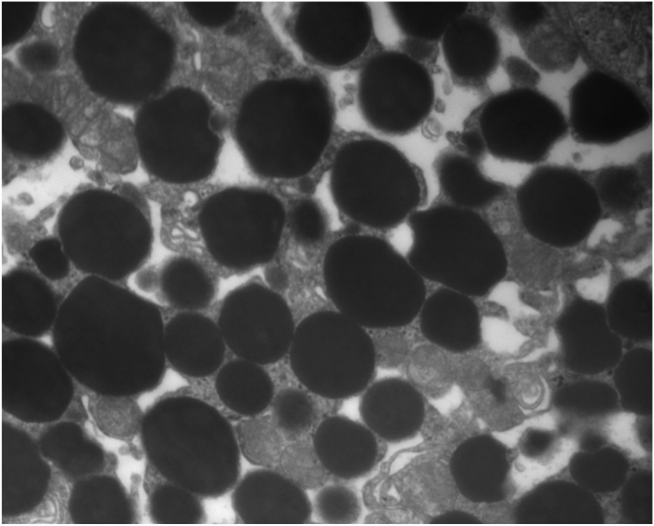
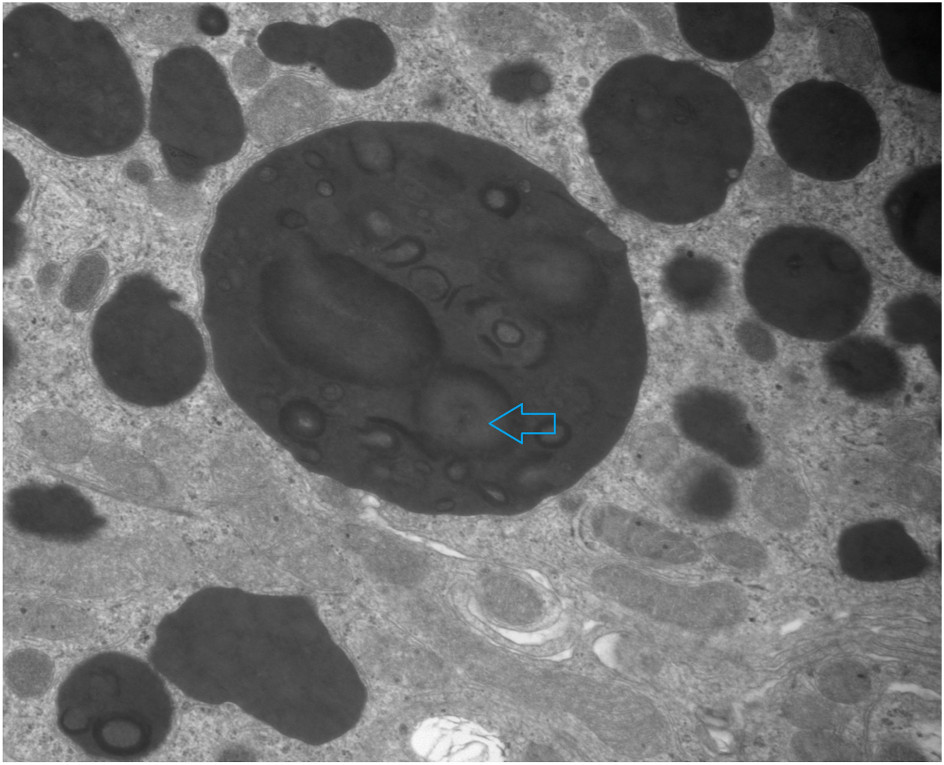
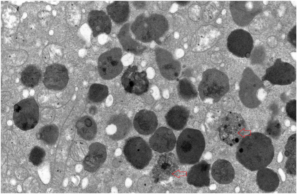
Ultrastructural findings: Two types of intracytoplasmic inclusions are present in proximal tubular epithelial cells.
These are intracytoplasmic, membrane-bound lysosomes containing homogenous electron-dense material, sometimes containing curvilinear/circumferential structures with central lucent area. In the second pattern, the lysosomes contain granular electron-dense materials, sometimes with admixed cellular debris, and are considered autophagolysosomes. These 2 appearances of lysosomes sometimes coexist in a single cell.
Lysozyme is produced by monocytes and increased in conditions with increase in myeloid cells.
Although elevated circulating lysozyme levels are important in the pathogenesis of lysozyme-associated nephropathy, the mechanism of the associated acute tubular injury is unclear. The increased lysozyme is taken up by proximal tubules by megalin-cubulin transport.
The presence of cell debris in some tubular cell autophagolysosomes suggests direct lysozyme-mediated acute tubular cell injury. It is also possible that impaired endolysosomal function is caused by direct toxicity of supersaturated lysozyme. Superimposed ischemic and/or toxic tubular damage may also contribute to lysozyme-mediated tubular injury.
Lysozyme-associated nephropathy should be distinguished from chromogranin-associated tubulopathy and noncrystalline light chain proximal tubulopathy. Immunohistochemical staining for lysozyme demonstrates diagnostic staining of the inclusions. Immunostain for chromogranin A is negative in lysozyme-associated nephropathy. Correlation with serum lysozyme level and urine protein electrophoresis demonstrating excess excretion of lysozyme are essential for establishing a diagnosis. In noncrystalline light chain proximal tubulopathy, immunofluorescence following pronase digestion shows strong monotypic kappa or occasionally lambda staining in proximal tubular epithelial cells, which is negative in lysozyme-associated nephropathy. Whether this staining in noncrystalline light chain proximal tubulopathy reflects trafficking of a monoclonal protein, or is causal in the acute tubular injury, is not definitely proven. In classic crystalline light chain proximal tubulopathy, in addition to clonal staining after pronase digestion, characteristic crystalline, rhomboid, or trapezoid inclusions are present by electron microscopy in proximal tubular cytoplasm.
The primary treatment for lysozyme nephropathy is supportive care for the acute tubular injury and treatment of the underlying disease. At present, there are no therapeutic interventions that specifically target lysozyme-mediated tubular injury. Prognosis is variable, depending on type of underlying hematologic malignancy and whether there is neoplasm in the kidney. Patients with direct involvement of the kidney by the underlying neoplasm have a worse prognosis. In one series with 8 months follow-up information, 8 of 30 patients (27%) died. Of the 4 patients with direct involvement of the kidney by neoplasm, 3 died and 1 was placed on palliative care. Treatment of the underlying neoplasm improved kidney function with increase in estimated glomerular filtration rate greater than 15 mL/min/1.73 m² in some patients in this series.
These links are current-reference entry points; the atlas text above preserves the source article wording. External links checked 2026-09-24.