Microscopy Images
Patients with malakoplakia usually present with fever, flank and/or loin pain, and a history of E. coli urinary tract infection. Malakoplakia may occur at any age, but most commonly presents in the fifth decade of life with a predilection for women over men (4:1). Patients may be immunosuppressed, with underlying conditions such as AIDS, diabetes, alcohol abuse, or another immunocompromised state. Bilateral kidney involvement occurs in less than half of cases and usually presents with acute kidney injury. Prognosis has markedly improved with use of fluoroquinolone drugs, with >90% patient survival.
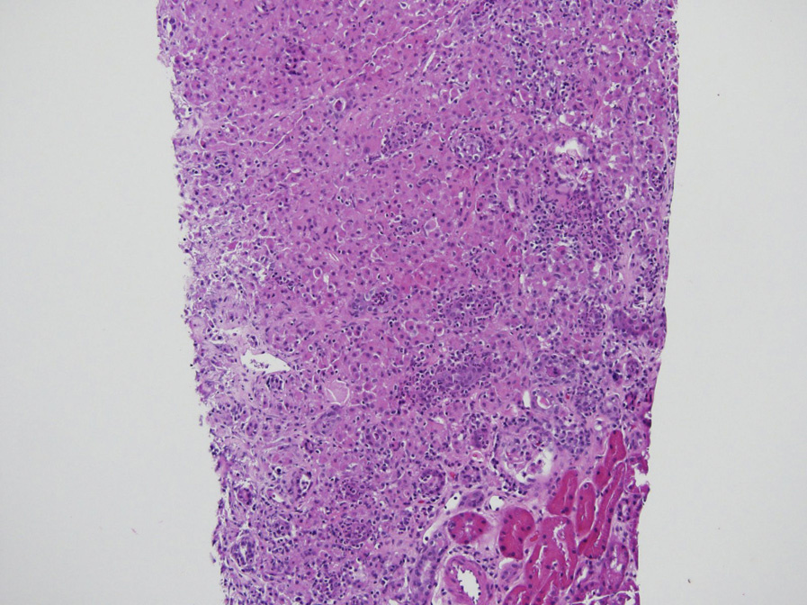
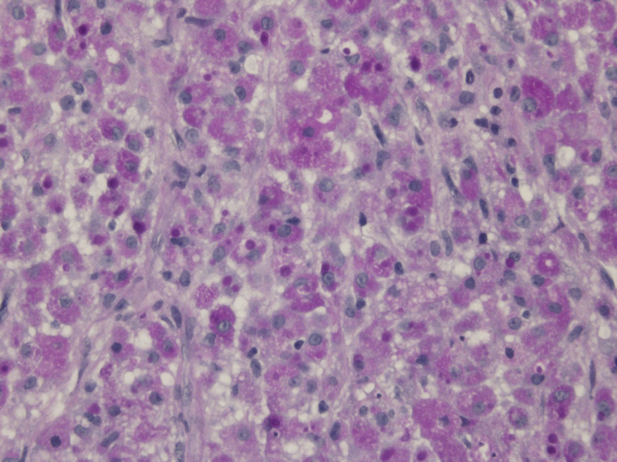
Light microscopy: Sheets of macrophages, forming mass-like lesion, with granular eosinophilic cytoplasm and characteristic inclusions of periodic acid– Schiff–positive Michaelis-Gutmann bodies, which also stain for calcium and iron.
Immunofluorescence microscopy: Noncontributory.
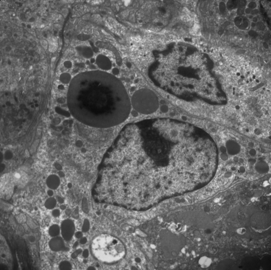
Electron microscopy: The macrophage intracytoplasmic Michaelis-Gutmann bodies have crystalline central core with peripheral lamellar rings of mineral deposits.
Macrophage inability to release lysosomal enzymes leads to decreased degradation of bacteria, most commonly E. coli. This defect in macrophage intracytoplasmic bactericidal function results in partially digested bacterial products that form a nidus for calcium and iron minerals.
Renal cell carcinoma can be distinguished from malakoplakia by positive cytokeratin staining in the absence of macrophage stains (for CD163 and CD68).
Xanthogranulomatous pyelonephritis has lipid-laden foamy macrophages without intracytoplasmic Michaelis-Gutmann bodies. Megalocytic interstitial nephritis has macrophages with granular eosinophilic cytoplasm, also without intracytoplasmic MichaelisGutmann bodies. Megalocytic interstitial nephritis is also closely linked to E. coli urinary tract infection and may represent an earlier stage of malakoplakia.