Microscopy Images
Lupus nephritis (LN) is defined as glomerular immune complex disease that occurs in patients who meet American College of Rheumatology criteria for diagnosis of systemic lupus erythematosus (SLE). SLE is a systemic autoimmune disease, most commonly involving the skin, kidneys, joints, heart, and serosal surfaces. Women are affected more than men (9:1), and SLE is more common in African Americans. Onset usually is from teenage years to the third decade of life, but SLE may manifest at any age. Kidney involvement is a major cause of morbidity and the most common cause of death in SLE patients.
The varying glomerular immune complex patterns are diagnosed according to the ISN/RPS classification. These include predominantly mesangial deposits (classes I, II), subendothelial deposits with endocapillary hypercellularity or prominent glomerular basement membrane (GBM) duplication and wire-loop lesions, often with necrotizing and crescentic lesions (classes III and IV, focal and diffuse LN), or membranous forms (class V).
Membranous LN (class V) is seen in up to 15% of biopsied SLE patients, who typically present with nephrotic syndrome, and can occur in combination with focal or diffuse LN (class III or IV); thus there may be additional clinical symptoms at presentation related to active lesions of focal or diffuse LN. In such cases, proteinuria may take longer to resolve with therapy than expected with proliferative LN alone.
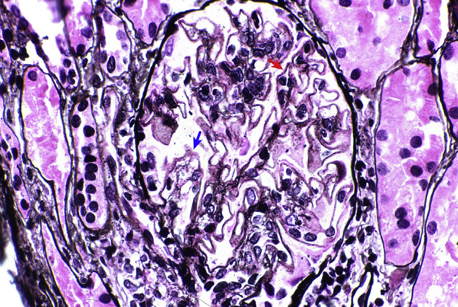
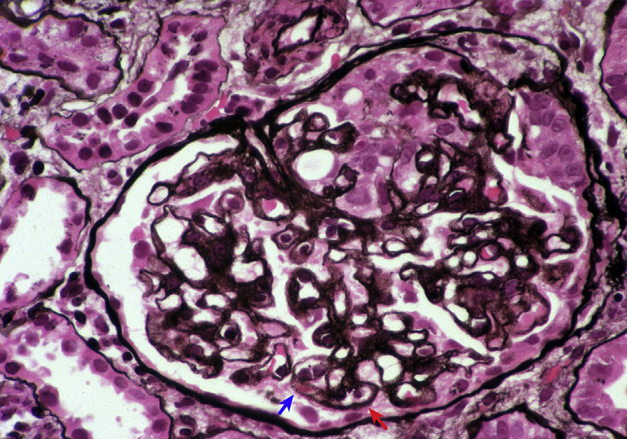
Light microscopy: GBMs are prominent with appearance of “holes” and “spikes” on silver stain in more advanced stages (in the earliest stages, they may not be apparent), accompanied by mesangial matrix expansion and increased cellularity. Additional lesions, including crescents, fibrinoid necrosis and/or endocapillary hypercellularity or segmental sclerosis related to scarring of active lesions, are present when membranous LN occurs in combination with proliferative LN forms (class III or IV). Segmental sclerosis of usual type related to podocyte injury associated with membranous nephropathy may be difficult to distinguish from scarring of active lesions. Wedgeshape fibrous adhesion of glomerular tuft to Bowman capsule favors scarring from a previously active lesion rather than usual type segmental sclerosis. Interstitial fibrosis may be seen with disease progression.
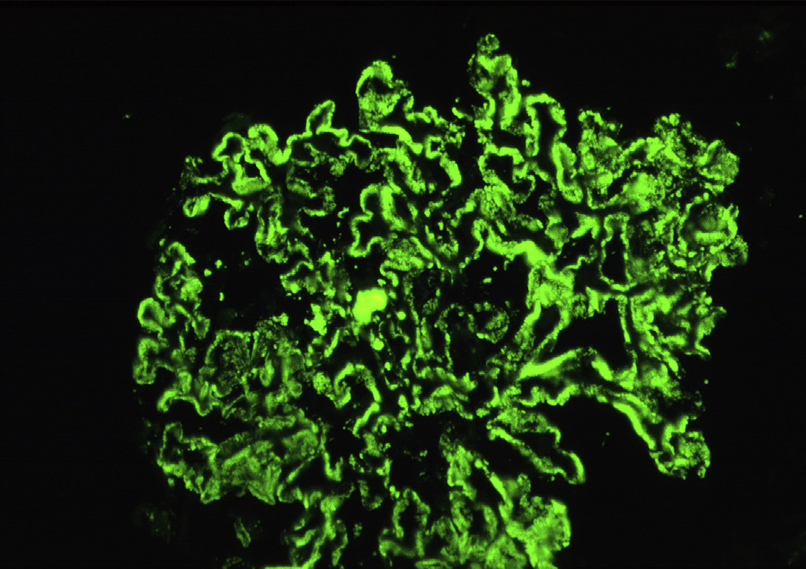
Immunofluorescence microscopy: More than half of glomerular capillary loops in more than half of glomeruli show granular staining of dominant or codominant IgG in a “full-house” polyclonal pattern.
Nuclear staining for IgG, reflecting positive ANA, may be present. Granular immune complex deposits rarely involve peritubular capillaries, particularly with staining for C1q. Granular tubular basement membrane and vascular deposits, either focal or diffuse, occur commonly, with variable lymphoplasmacytic infiltrates.
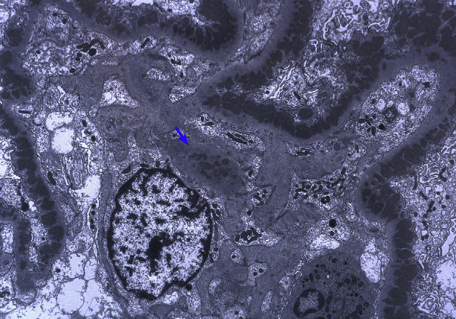
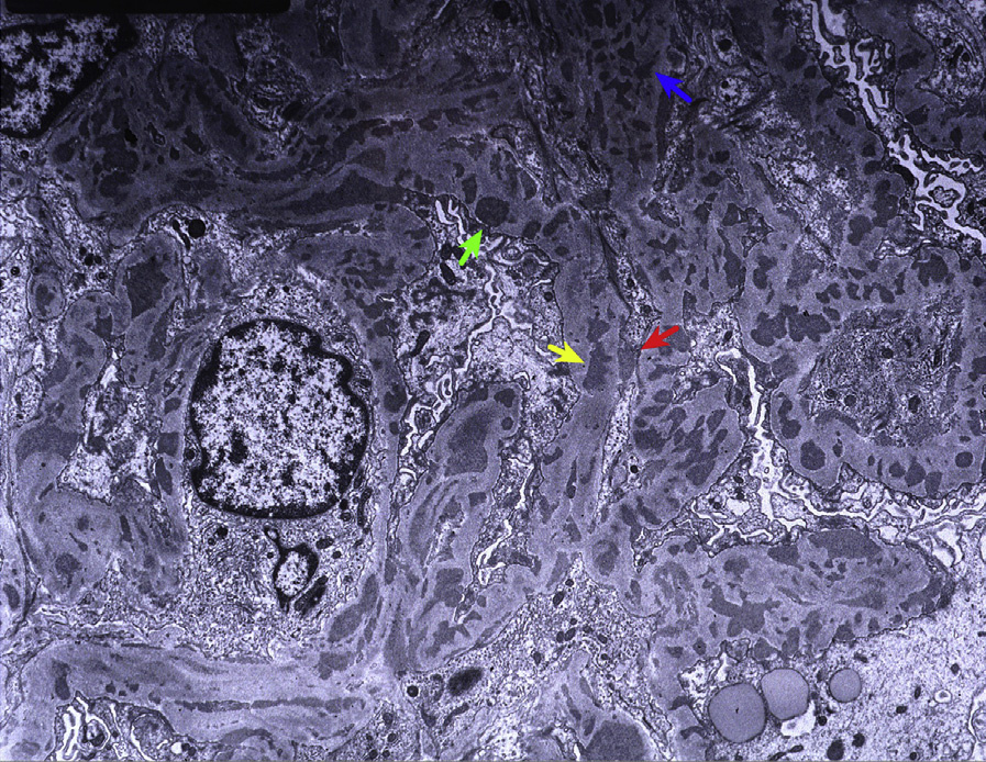
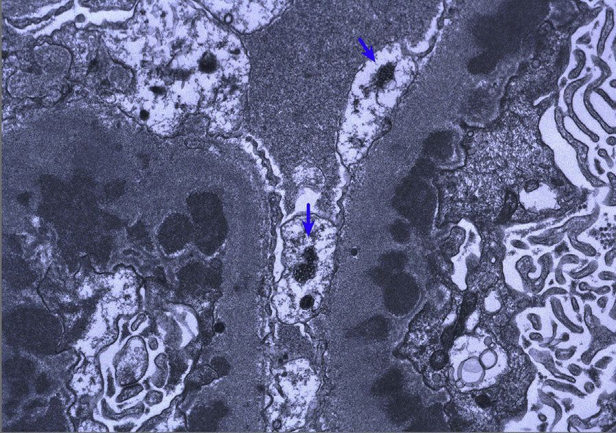
Electron microscopy: There are scattered to frequent subepithelial deposits involving more than half of capillary loops of more than half of glomeruli with mesangial deposits and there may be occasional subendothelial deposits. Reticular (also known as tubuloreticular) aggregates are seen in endothelial cell cytoplasm.
There are genetic predispositions to SLE. Inadequate clearance of nuclei from cells undergoing apoptosis (triggered by various environmental insults, including ultraviolet radiation) leads to excess nuclear antigens which, combined with additional abnormalities in B and T lymphocytes, ultimately results in autoantibody formation. The activation of Toll-like receptors and dendritic cells and high interferon a levels are additional elements postulated to perpetuate this process.
Autoantibodies develop against a variety of nuclear materials, including DNA, RNA, and various histone and nonhistone proteins.
Primary membranous nephropathy typically does not have full-house immunofluorescence staining, nor significant mesangial deposits, subendothelial deposits, or reticular aggregates by electron microscopy as seen in LN. Up to 70% of primary membranous nephropathy cases show PLA2R-positive immunostaining, which rarely occurs in LN.
Other causes of secondary membranous nephropathy, including mixed connective tissue disease and lupus-like illnesses, need to be distinguished clinically.